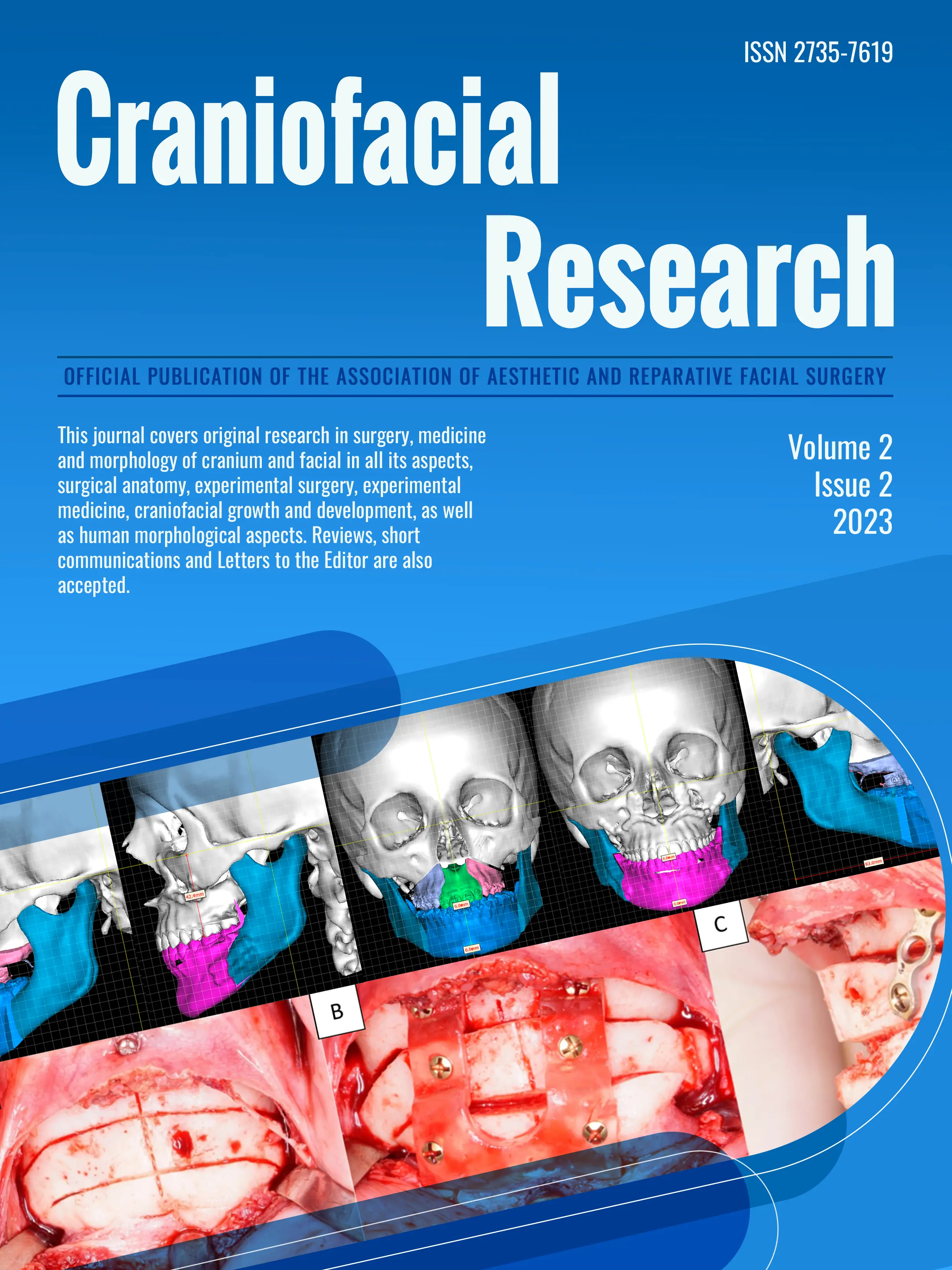
Welcome to Craniofacial Research
Craniofacial Research is a quarterly publication that aims to disseminate original research and research methods related to surgery, medicine, and cranial and facial morphology. It welcomes submissions on a wide range of topics, including surgical anatomy, experimental surgery, experimental medicine, craniofacial growth and development, and human morphological aspects. In addition to research articles, the journal also accepts reviews, short communications, and letters to the editor. All articles are subjected to rigorous evaluation by independent expert reviewers.
Latest published issues

Most visited post
1. Analysis of biomimetic strategies for surface conditioning of dental implants: a literature review
It has been seen that enhancing the characteristics of the surface of an implant with bioactive agents that seek to mimic the chemical and biological nature of native bone can influence osseointegration. This article seeks to describe biomimetic treatment strategies for dental implant surfaces that favor the bioactivity of implant materials. The search was performed in PUBMED and the Cochrane Library with the terms "biomimetic dental implants" or "biomimetic implants" obtaining 110 results. After analyzing each reference, it was determined that 11 articles coincide with the objective of this review, they provide data from ex- perimental studies, reviews, and clinical cases on different biomimetic strategies for surface treatment. Within the surface conditioning strategies with chemical substances, it is described that sandblasting and acid etching favor biocompatibility, cell differentiation and bone formation. Also, the use of coatings that allow the composition of the implant surface to approach the na- tural characteristics of bone tissue is favorably described. On the other hand, based on a biomimetic approach, implant surface coatings with different biomolecules such as growth factors, extracellular matrix proteins, peptides and drugs are described, with the constant objective of promoting osseointegration. Although studies indicate that implant surface treatments favor the osseointegration process, it is necessary to investigate mostly the use of biomolecules that combine the implant surface with the host environment.
2. Arthrocentesis of the temporomandibular joint with triangulation technique: technical data
Arthrocentesis is a minimally invasive treatment indicated for internal TMJ (temporomandibular joint) disorders. However, it requires the expertise of a specialist and presents a significant technical difficulty due to the three-dimen- sional small size of the joint. In this technical note we describe the insertion of the second arthrocentesis needle based on the triangulation technique described by McCain for arthroscopy.
3. Surgical management and follow-up of custom implant treated orbito-zygomatic fracture: case report and literature review
Orbital floor fractures are common injuries in the maxillofacial region, typically caused by blunt trau- ma. Clinical manifestations involve facial asymmetry, enophthalmos, diplopia, and restricted ocular motility. These fractures can be treated by either conservative or surgical approaches. We present the case of a 59-year- old patient with a history of orbitozygomatic fracture, resulting from a car accident, treated using a custom- made polyetheretherketone (PEEK) implant at the Hos- pital Militar de Santiago. Using virtual planning software and 3D printing technology, a PEEK implant was designed and positioned via a transconjunctival approach. Long-term follow-up showed favorable evolution without loss of visual acuity and evidence of bone regeneration at five years. This case highlights the advantages of PEEK in orbital reconstruction, including its adaptability and biocompatibility, while acknowledging challenges such as cost and infection risk, underscoring its potential in modern maxillofacial surgery.
4. Extreme facial rejuvenation, the combination of orthognathic surgery and cervico-facial rhytidectomy
Multiple adjunctive procedures are performed with orthognathic surgery to enhance aesthetic facial results, especially in those cases where skeletal movements alone do not satisfy the aesthetic demands of the patient. The objective of this study is demonstrate the benefits and outcomes of performing orthognathic surgery and cervicofacial rhytidectomy simultaneously to achieve extreme facial rejuvenation. Two clinical cases with a diagnosis of cervicofacial rhytidosis and dentoskeletal anomalies are shown, the first case with retrogeny and superior dermatocalasia and the second case with class III dentofacial anomaly and retrogeny, which were treated with the combination of orthognathic surgery and cervicofacial rhytidectomy in the same operative act. Facial aesthetic surgery has evolved by recognizing that changes in hard tissues affect soft tissues, however its isolated resolution does not meet the final aesthetic expectations of the patient in soft tissues. That is why there is a need to combine simultaneous procedures to meet the needs for which the patient comes and maintain the results over time. All patients who decide to undergo surgery for aesthetic reasons must also improve function. And due to the increase in operative time that carries out both surgeries simultaneously must be performed by a well-trained maxillofacial surgeon.
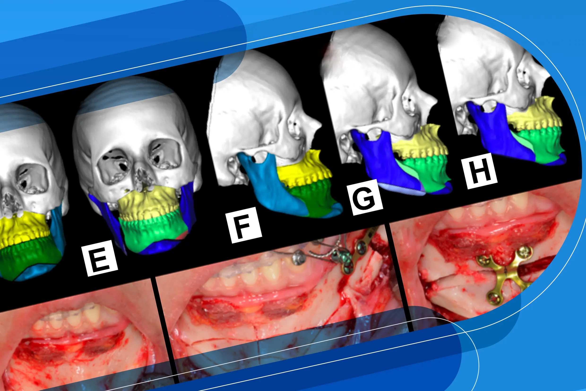
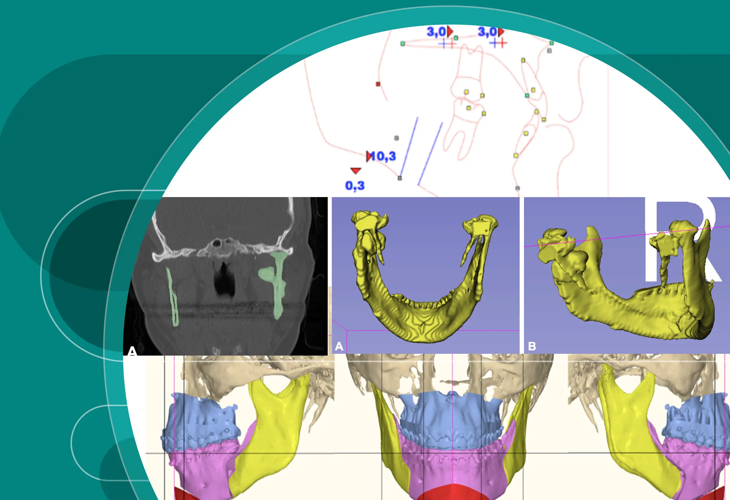
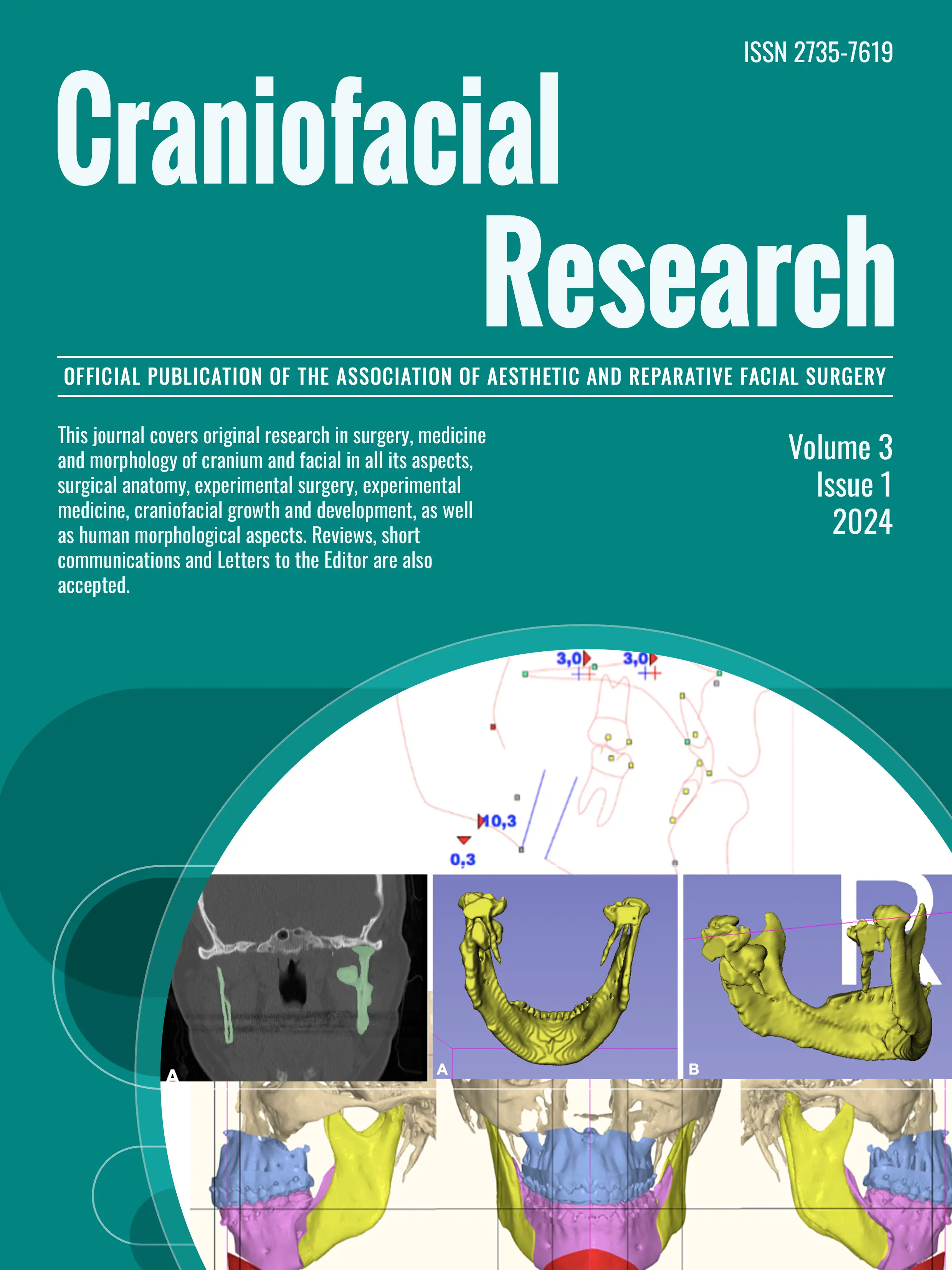
5. Stepped genioplasty using cutting and positioning guide: A case report
Genioplasty is a procedure used to modify the natural anatomy of the chin, based on an osteotomy of the lower edge of the jaw that allows three-dimensional repositioning of the chin. Currently, various techniques are described to perform genioplasty, being the staggered technique, a modified technique used for elusive chins with vertical excess of the lower third. A genioplasty, virtually previously planned, was performed with a stepped technique using a cutting and positioning guide in an 18- year-old patient with a history of class II dentofacial dysmorphosis that affected both functionally and aesthetically the lower third. Genioplasty with the stepped technique allows addressing the vertical excess of the chin, both at the level of the symphysis, allowing to manage the projection of the chin and deepening of the labiomental fold, achieving smooth transitions between the symphysis and the ramus of the mandible in comparison with other techniques. conventional. The use of cutting and positioning guidance allowed to obtain previously planned intraoperative osteotomies, obtaining virtually predictable and complication-free planned aesthetic results.
6. Simultaneous use of double Struts for unsupported noses: technical note
Rhinoplasty is the most practiced facial aesthetic procedure in the world and has generated enormous literature on the matter. The objective of this study is to describe the use of the simultaneous double strut technique for unsupported noses. Currently the anterior strut does not need a great length, nor does it need to be fixed to the SNA, just as it will not be in charge of providing projection to the nasal tip, it is enough to fix it to the medial cruras to provide greater support to the nasal tip. the technique Teo strut, is in charge of giving projection as well as the support to the nasal tip. The Teo Strut technique possible to provide support and stability to the nasal projection, rotation of the nasal tip, and adequate points of light and shadows in the different polygons, maintaining principles of preservation in some cases, thus reduci
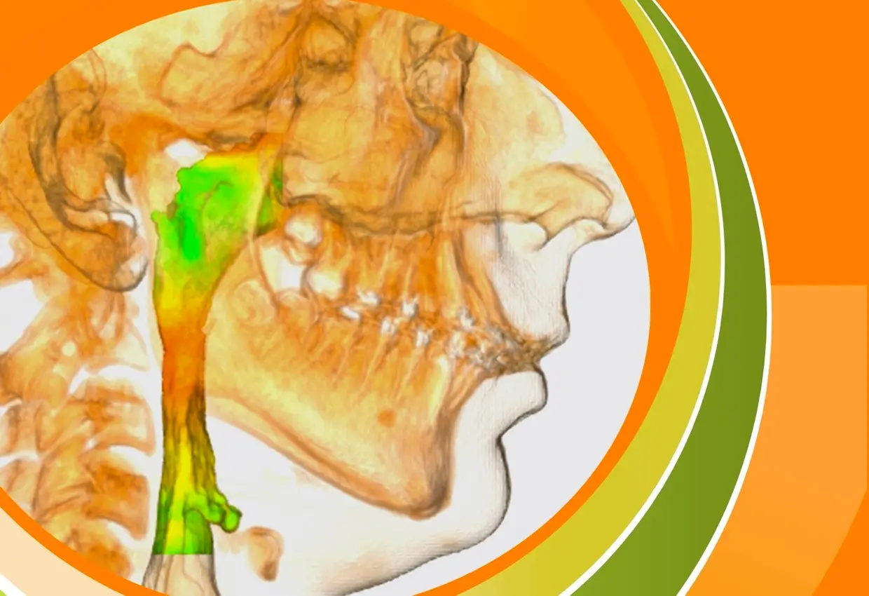
7. Relationship between maxillo-mandibular rotation and airway volume in subjects with maxillo-mandibular prognathism
The vertical growth of the mandible is related to the degree of inclination of the mandibular plane and the contour of the mandibular angle. These characteristics are related to the upper airway and vary depending on the skeletal class and the mandibular vertical pattern. The aim of this study is to describe and compare the inclination and angulation of the maxillomandibular plane in class III subjects and to establish the relationship with the area and volume of the airway. A cross-sectional study was performed to evaluate the area and volume of the airway and itsrelationship with the maxillomandibular plane in class III skeletal subjects who are candidates for orthognathic surgery, using cone beam computed tomography. A total of 86 CIII subjects with mandibular prognathism who were candidates for orthognathic surgery were analyzed. They ranged in age from 18 to 59 years (29 ± 10.1). 46 subjects were male (55.55%) and 40 were female (44.45%). 57 subjects presented an open angulation and 29 subjects presented a closed angulation. Subjects with an open angulation presented a hyperdivergent pattern. Whereas subjects with a closed angulation presented a greater minimum area (p<0.01), maximum area (p<0.07) and to- tal volume (p<0.03) than subjects with an open angulation. We can conclude that in skeletal CIII subjects with mandibular prognathism who present open angulation, it is necessary to evaluate the mandibular inclination together with the minimum area and total volume of the upper airway.
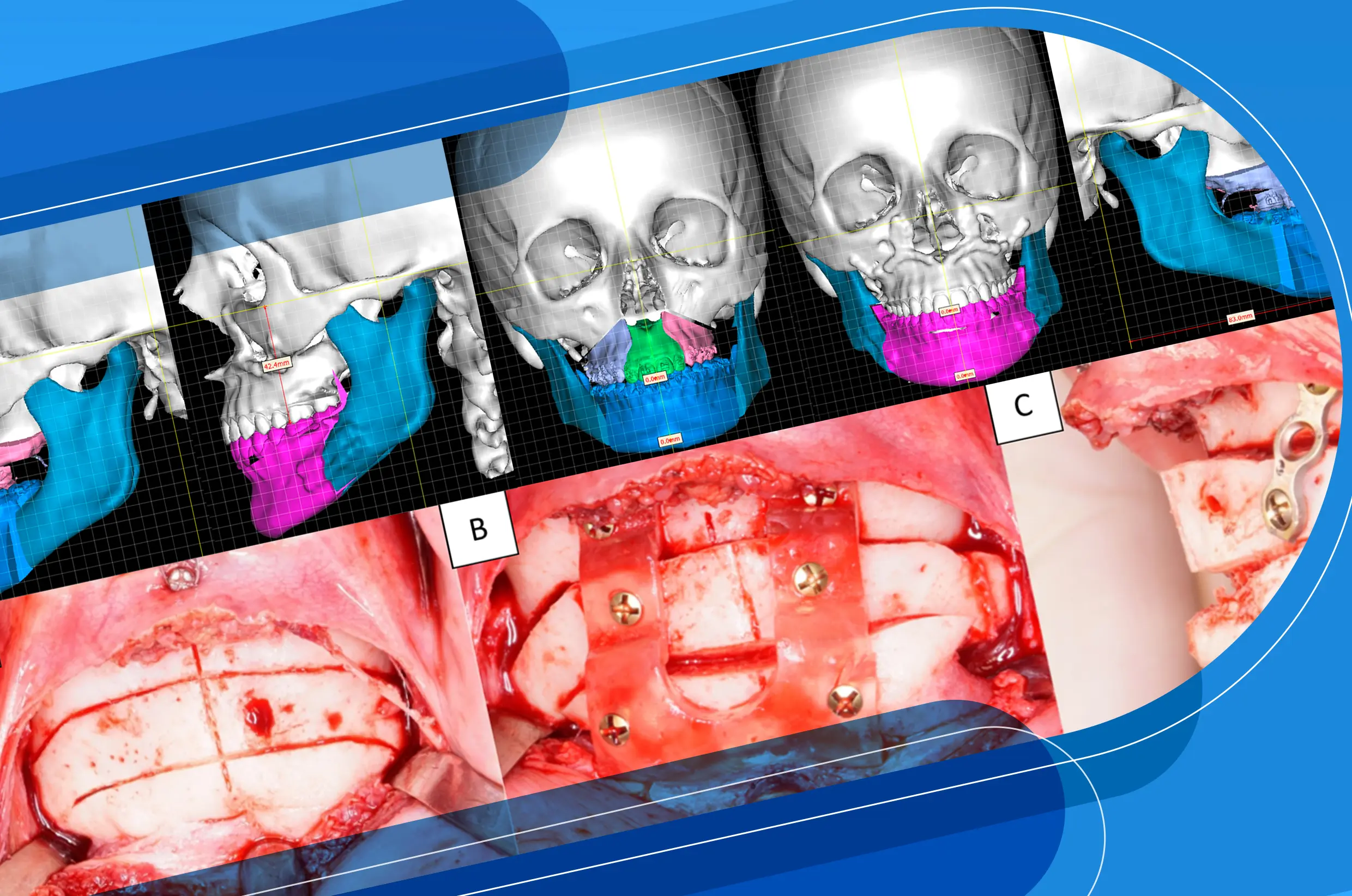
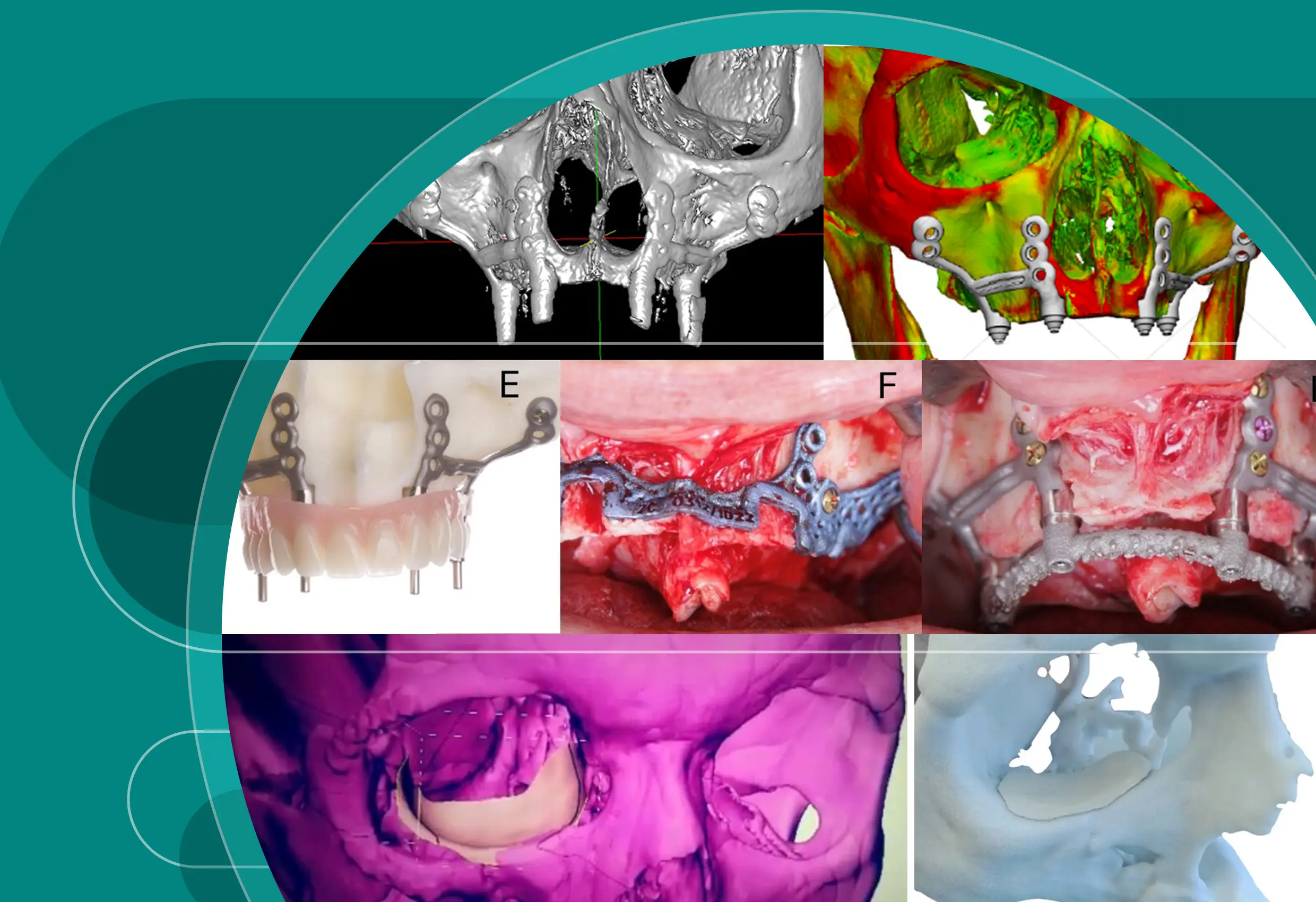
8. Extraoral exposure of mandibular reconstruction plate
Maxillofacial reconstruction systems have presented an incredible advance in terms of implementation techniques and properties. These systems have been adapted throughout history, through medical and orthopedic principles, exceeding minimum requirements in relation to their resistance (to provide functional stability), ductility, and above all, biocompatibility. In this way, experimental studies in both biomechanics and pathophysiology of osseointegration have given these system high reliability and prognostic accuracy. However, there are limitations and requirements to be considered, in order to avoid complications such as fracture of the material, loss of stability of the rigid fixation, infection, both intra and extra oral exposure of the reconstruction plate, among others. The purpose of this article is to report a case of extra oral exposure of a mandibular reconstruction osteosynthesis plate in a patient undergoing hemimadibulectomy after severe mandibular osteomyelitis, presenting the diagnosis, management and treatment performed by the team of specialists.
9. Profunda femoris artery flap as an option in tongue reconstruction. A technical note
Reconstruction of the tongue following ablative surgery is a well-documented technique that shows significant improvement in swallowing, speech, and quality of life. Free flaps, such as the anterolateral thigh, forearm, rectus abdominis, and latissimus dorsi flaps, are the most commonly used in tongue reconstruction. Additionally, in recent years, the profunda femoris artery perforator flap (PAP flap), discovered by Argentine surgeon Claudio Angrigiani in 2000, has gained recognition in head and neck reconstruction, particularly for tongue reconstruction. This article aims to present a technical note focused on the application of the PAP flap in tongue reconstruction.
10. Intravascular papillary endothelial hyperplasia in the lower lip: A case report
Intravascular papillary endothelial hyperplasia or Masson's tumor is a benign, non-neoplastic lesion that can affect the torax, fingers, head, and neck. Its presence in the oral cavity is unusual; however, there are reports in the literature. A female patient in the 4th decade of life who attended the Oral and Maxillofacial Surgery Department of the Universidad Central de Ve- nezuela, with a purplish lesion on the lower lip of two months of evolution associated with masticatory trauma is presented. An excisional biopsy was performed, and a histopathological study was performed. The wall of blood vessels made up of endothelial cells showing intraluminal papillary growth was observed. The papillary structures are lined by hyperplastic endothelial cells in the vascular lumen. The definitive diagnosis was intravascular papillary endothelial hyperplasia. Due to the great resemblance to other vascular lesions, it is essential to perform a histopathological study of suspicious lesions to rule them out.